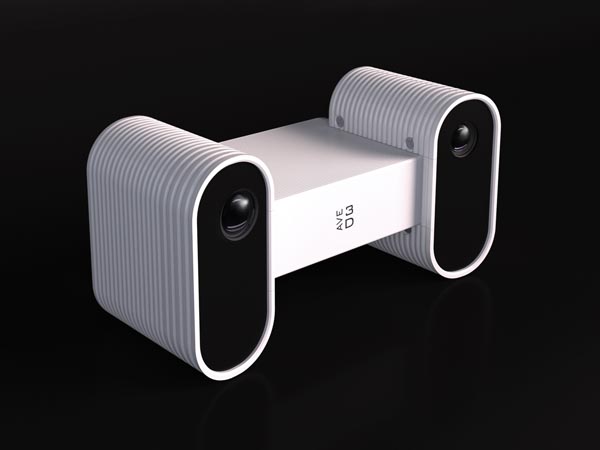
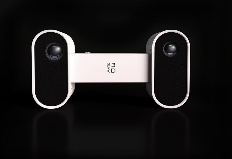
AVE 3D HEAL
WORLD’S FIRST 3D SCANNER FOR DIAGNOSIS AND OBJECTIFICATION OF PATIENTS IN MOVEMENT
Innovative 3D motion scanning technology which unveils new possibilities in orthopedics, neurology, gaming, sports, and aesthetic medicine, enabling precise analysis and monitoring of dynamic processe

MEASURABLE 3D MOVIE WITH MOTION
A REVOLUTIONARY SYSTEM FOR DIAGNOSTICS AND DATA OBJECTIFICATION IN PRE AND POST SURGICAL TREATMENT
Medicine and rehabilitation
We register a 3d movie in just a few seconds. Acquired data in real time can be used to reproduce body movement for the diagnosis and treatment of various orthopedic, neurological, and other conditions. They can also be used to help design prosthetics and for rehabilitation after injury.
SCANNER
Our 3D scanner as Your shield against patients claims
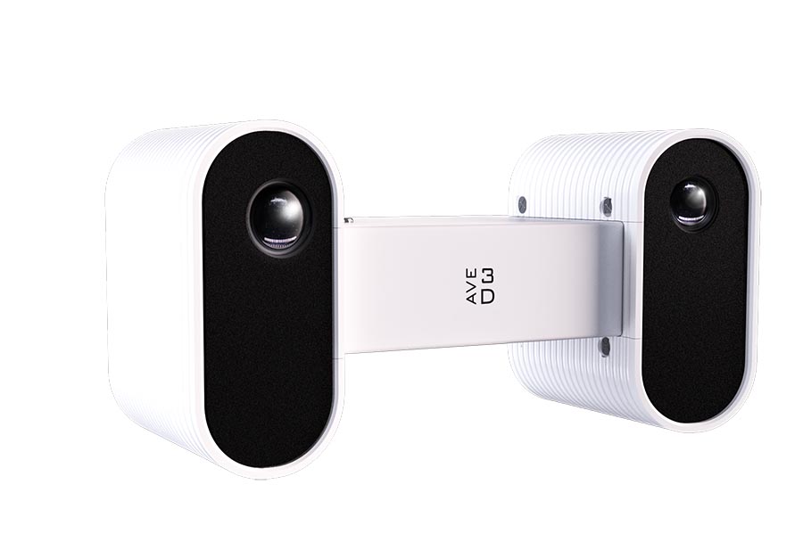
AVE 3D Heal is the world’s first scanner that allows 3D dynamic scanning of the geometry of the body’s limbs, with a particular focus on the feet and wrists. Unlike traditional medical imaging, the 3D scanner works non-invasively, eliminating the need for X-rays. As a result, doctors and physiotherapists receive detailed information about the shape, anatomical structure, and possible deformities and swellings, which is important information for issuing a precise diagnosis on the subject of the patient’s condition. The biggest advantage of the developed solution is the possibility of spatial imaging of the patient in motion, and thus it allows generating detailed documentation including three-dimensional examination results, which can be used for analysis, comparisons of treatment progress, as well as for research and scientific purposes.
DEDICATED TO MEDICAL PROFESSIONALS
3D GONIOMETER – MEASURE, COMPARE
AND CONTROL
Comprehensive assessment:
Dynamic scanning enables the capture of a wider range of data, including information about joint mobility and muscle function, which can provide a more comprehensive assessment of the patient’s condition.
3D models from each second
Our software is using pattented technology called ONE SHOT. It enables to export digital models of particulart parts of the body. You can freeze-frame registered 3d movie at any second and export captured earlier 3D data from particular position in movement. We can export data in such formats as *.STL ; *.PCS ; *.CLOUDDATA
Improved visualization:
The 3D movie generated from the dynamic scan can be manipulated and viewed from different angles, providing a more detailed and interactive visualization of the scanned area.
Automatic report generation
AESTHETIC MEDICINE
Capabilities of our device for facial motion scanning in aesthetic medicine
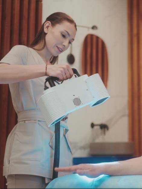
The Ave 3D Heal scanner allows for precise capturing of even subtle changes in facial expressions in real time, which allows for in-depth analysis of muscle dynamics and their impact on facial appearance.
This knowledge is extremely valuable for aesthetic medicine doctors, as it allow for accurate diagnosis of the causes of aesthetic problems, such as wrinkles, nasolabial folds, drooping eyelids, or facial asymmetry. Based on the 3D scan, it is possible to precisely determine the areas that require correction and select the appropriate techniques and preparations to achieve optimal results.
BENEFITS OF USING AVE 3D
The 3D scanner brings numerous benefits to professionals as well as patients. Its most important aspects include:
Shield for patient claims
The 3D scanner provides accurate and measurable information about the anatomical structure of the extremities, enabling doctors to effectively diagnose conditions
Patient safety
The elimination of X-rays makes the 3D scanner a safe solution, especially for children and people who are sensitive to radiation.
Documentation and control
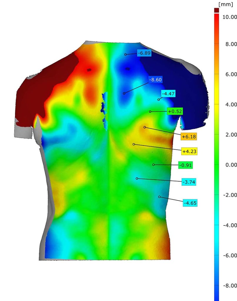
With Ave3D Heal 3D scanner, it is possible to document the 3D results of the limb examination in detail, thanks to reports created by the software. This documentation provides a valuable record that can be used for analysis, comparisons, and as a reference for possible future studies.User scans in motion and afterwards can export a digital model from particular second of that movie
Archiving for Research and Scientific Purposes
Precise 3D documentation from the 3D scanner can be used for research and scientific purposes by future specialists. Archiving the results allows for analysis of the data in the long term, which can contribute to advances in the field of orthopedics.
Facilitated Interdisciplinary Communication
Three-dimensional movie provide clear and understandable data, facilitating communication between different medical specialists. This, in turn, contributes to reducing errors in diagnoses.
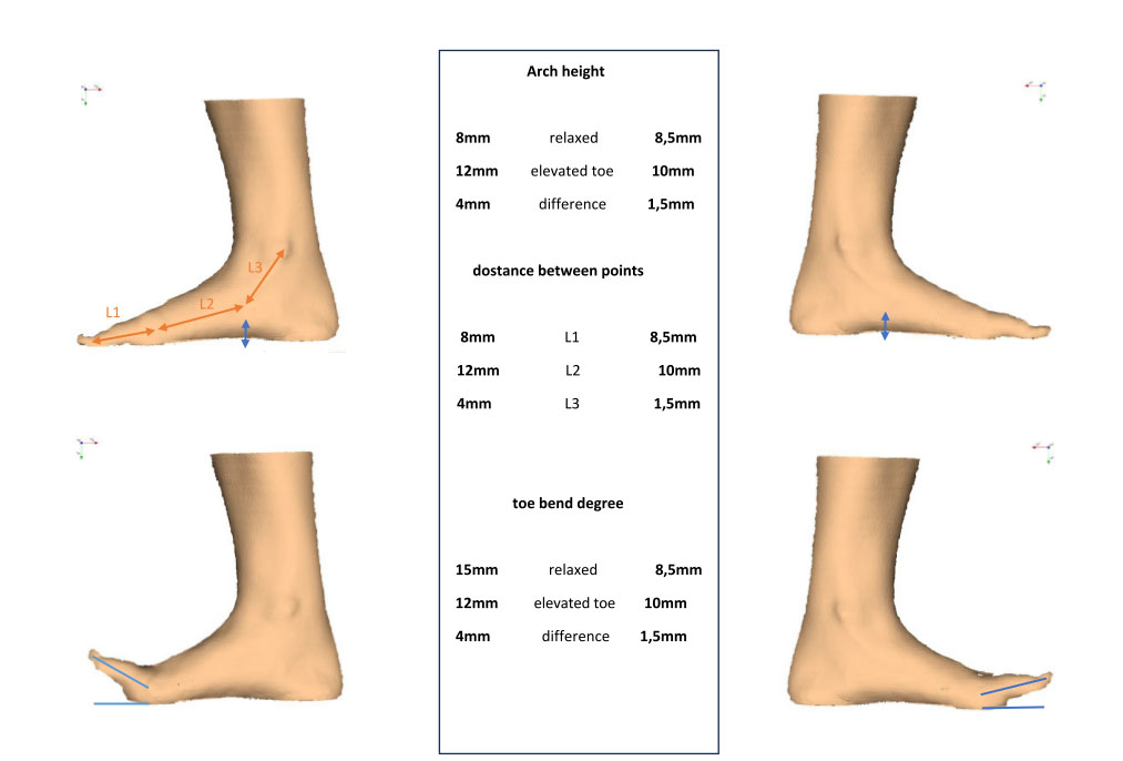
3D Goniometer – measure and compare
The 3D scanner offers highly precise measurements of the extremities' anatomical structure. This detailed information empowers doctors to diagnose conditions effectively. You Can compare both side and export 3d models afterwords.
